Your Weekend Read: Rediscovering the Diagnostic Potential of Vectorcardiograms - A Deep Dive for Medical Professionals
For professionals (mind our French): we're adding a new dimension to EKGs to better diagnose an aching heart.
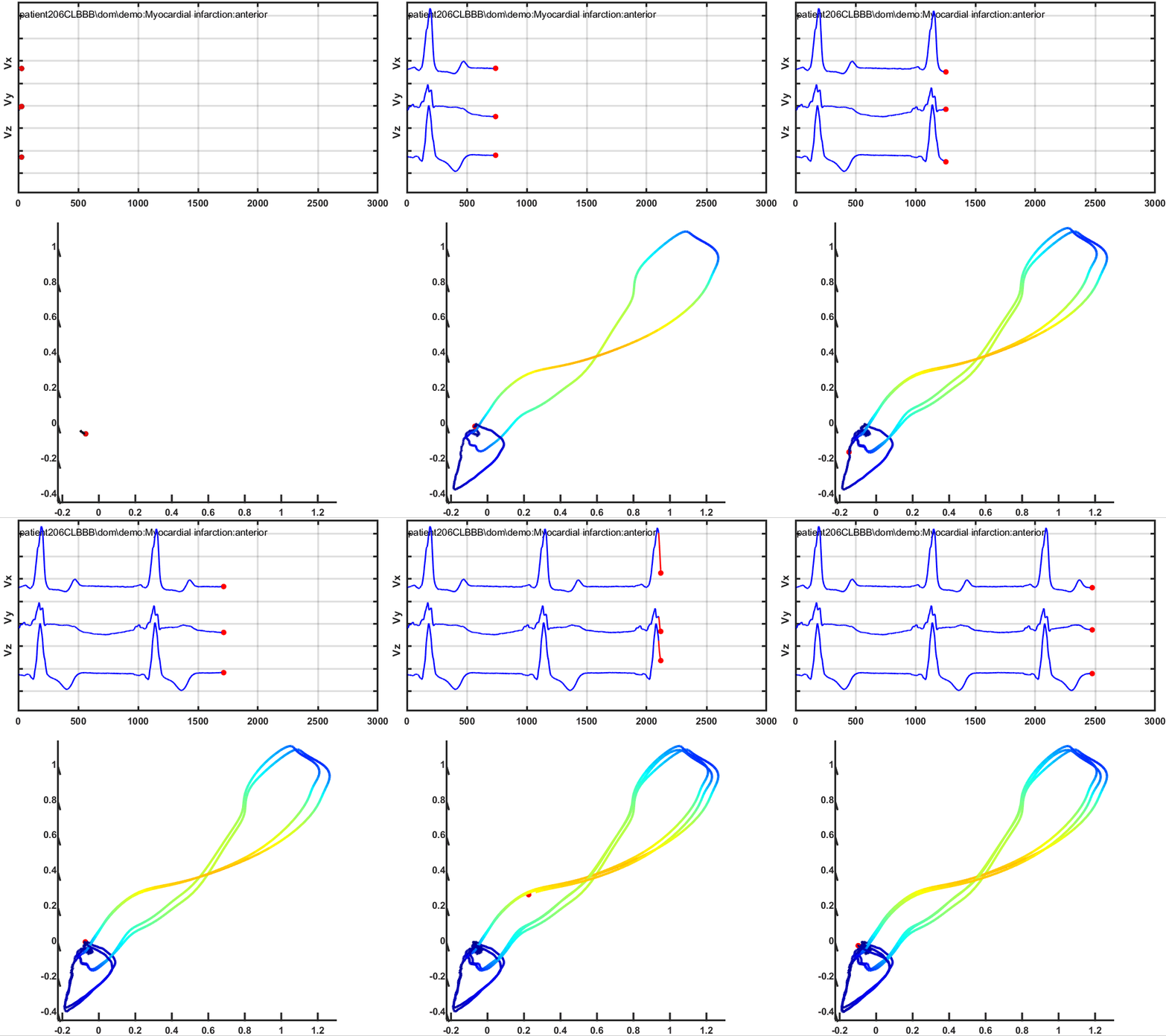
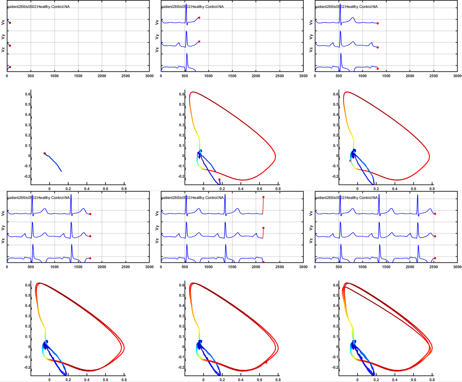
The Vectorcardiogram (VCG) is an important tool for cardiological diagnosis. Unlike a conventional Electrocardiogram (ECG), the VCG offers a 3D spatial representation of the electromotive forces generated during cardiac activity. This is done by analyzing these forces in three spatial planes - horizontal, frontal, and sagittal. The process involves the formation of an electric dipole during ventricular depolarization and the resulting dipole of cardiac electrical activity is represented by a vector, providing detailed information about the heart's electrical activity.
The VCG has been found to be superior to the ECG in certain specific situations such as evaluation of electrically inactive areas, identifying and locating ventricular pre-excitation, differential diagnosis of patterns varying from normal of electrical axis deviation, evaluating Brugada syndrome, and assessing severity of certain enlargements.
Vectors, which are units of measurement having direction, magnitude, and intensity, are crucial to the VCG. These vectors represent the dipole of depolarization and repolarization, and their loops represent the position of all instantaneous vectors during cardiac repolarization, providing different loops for the P, QRS, T and U waves.

The VCG uses Frank's system of corrected orthogonal leads that determine the three spatial planes. The term 'orthogonal' refers to the fact that the axes of the three planes are perpendicular to each other. Corrections are made for any deficient homogeneity of the electric field surrounding the heart.
The use of VCG has seen a resurgence in the 21st century with the advent of computerized vectorcardiography, enhancing processing and recording methods. While not a routine tool at most medical centers, it is invaluable in education, research, and for better morphological interpretation of the heart's electrical phenomena.
The following are the advantages of a vectorcardiogram (VCG) can be summarized as follows:
1. Three-dimensional Insights: A VCG provides a three-dimensional perspective of the heart's electrical activity, allowing more accurate spatial orientation and magnitude measurement of electrical vectors at any moment.
2. Improved Diagnostic Sensitivity: VCGs offer better sensitivity in detecting atrial enlargements and left ventricular enlargement (LVE). Studies have found that VCGs can diagnose around 50% of LVE cases, with a lower rate of false positives compared to ECGs.
3. Informative in Complex Cases: VCGs can be beneficial in cases of suspicion of electrically inactive areas in the septal or anteroseptal wall of the left ventricle, especially when systolic type LVE is present.
4. Stronger Correlation with Echocardiogram: VCGs correlate more effectively with echocardiograms in determining left ventricular mass, and can outperform both ECGs and echocardiograms in diagnosing chamber enlargement associated with electrically inactive areas.
5. Superior in Acute Myocardial Infarction (AMI): VCGs exhibit greater diagnostic sensitivity than ECGs in cases of AMI, particularly when associated with left anterior fascicular block (LAFB).
6. High Sensitivity and Specificity in Diagnosing Strict Dorsal AMI: VCGs can differentiate strict dorsal AMI from other conditions that also show prominent anterior forces, improving diagnostic accuracy.
7. More Sensitive in Diagnosing Multiple Infarctions: VCGs have shown higher sensitivity in diagnosing multiple infarctions, especially those associated with LAFB.
8. Increased Accuracy in Inferior Infarction Diagnosis: VCGs are generally more accurate than ECGs in diagnosing inferior infarctions, although some studies disagree.
9. Exceptional in Detecting Left Septal Fascicular Block (LSFB): VCGs are superior to ECGs in diagnosing LSFB, which is often underrecognized in certain medical literature.
10. Accurate in Identifying and Locating Anomalous Bundle: VCGs can accurately identify and locate the anomalous bundle in pre-excitation of the Wolff-Parkinson-White Syndrome.
11. Efficient in Diagnosing End Conduction Delays: VCGs are more sensitive and specific than ECGs in diagnosing end conduction delays in the fascicles of the right branch.
12. Great Value in the Analysis of Septal Percutaneous Ablation: VCGs are valuable in analyzing the electrical modifications resulting from septal percutaneous ablation of the obstructive form of severe hypertrophic cardiomyopathy.
Remember that while VCGs have many advantages, their use should be considered alongside other diagnostic tools for a comprehensive understanding of a patient's heart health.
VCG is a versatile tool that provides a more complete view of the heart's electrical activity, and can offer more accurate diagnoses in many cases than the EKG. It's particularly useful in diagnosing heart enlargements, heart attacks, and certain types of blockages, as well as monitoring high-risk procedures and evaluating the effects of treatment. It simplifies the process of obtaining precise graphs and measurements, including T wave dimensions and loop areas. Offering higher accuracy and faster processing than traditional methods, VCG continues to evolve. Despite similar diagnostic capacities to ECG, VCG's didactic utility and cost-effectiveness make it valuable, particularly where electrocardiographic recordings are ambiguous.


